Introducing… Biological Electron Microscopy Techniques!
Most students first hear of electron microscopy (EM) in their introductory biology course; it’s buried amongst a few other forms of microscopy, and perhaps even other techniques. At most, they’re expected to memorize a simple definition (it’s a technique involving a microscope that uses accelerated electrons as a source of light) and write it down on a quiz. But electron microscopy is a scientific art that takes multiple semesters and years of practice to truly master. To give students a taste of what it’s really like, the BIT Program is launching a new course next semester, Introduction to Biological Electron Microscopy Techniques.

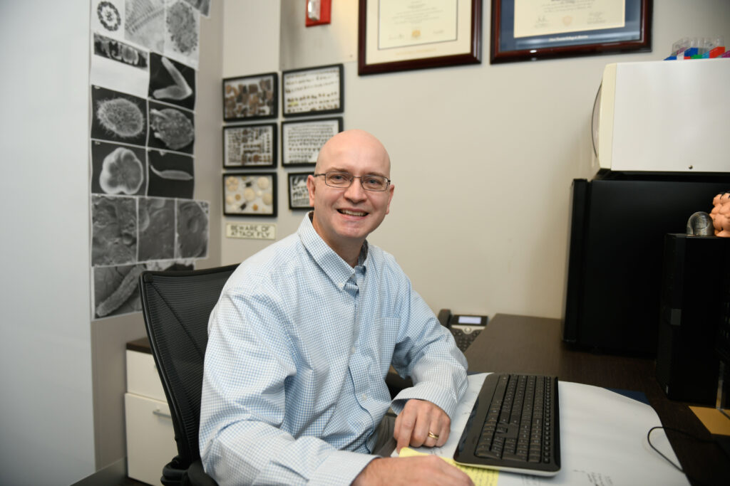
The course will be taught by Dr. Aaron Bell from the Analytical Instrumentation Facility (AIF). The AIF contains a multitude of state-of-the-art instruments to assist labs in their research, from X-ray diffraction to nanoindentation to, you guessed it, biological electron microscopy. Dr. Bell first started working with electron microscopy in 1997, when he was completing an MS in biology with an emphasis on EM at Hofstra University. He then went on to a PhD at the Albert Einstein College of Medicine, where he wasn’t able to work with EM as much as he wanted to. “I really wanted to go back to it,” he said, “so I accepted a position as the Head of EM at the New York Blood Center in 2011.” He moved to the AIF in 2019: “There aren’t a lot of jobs in the field, so I got really lucky.”
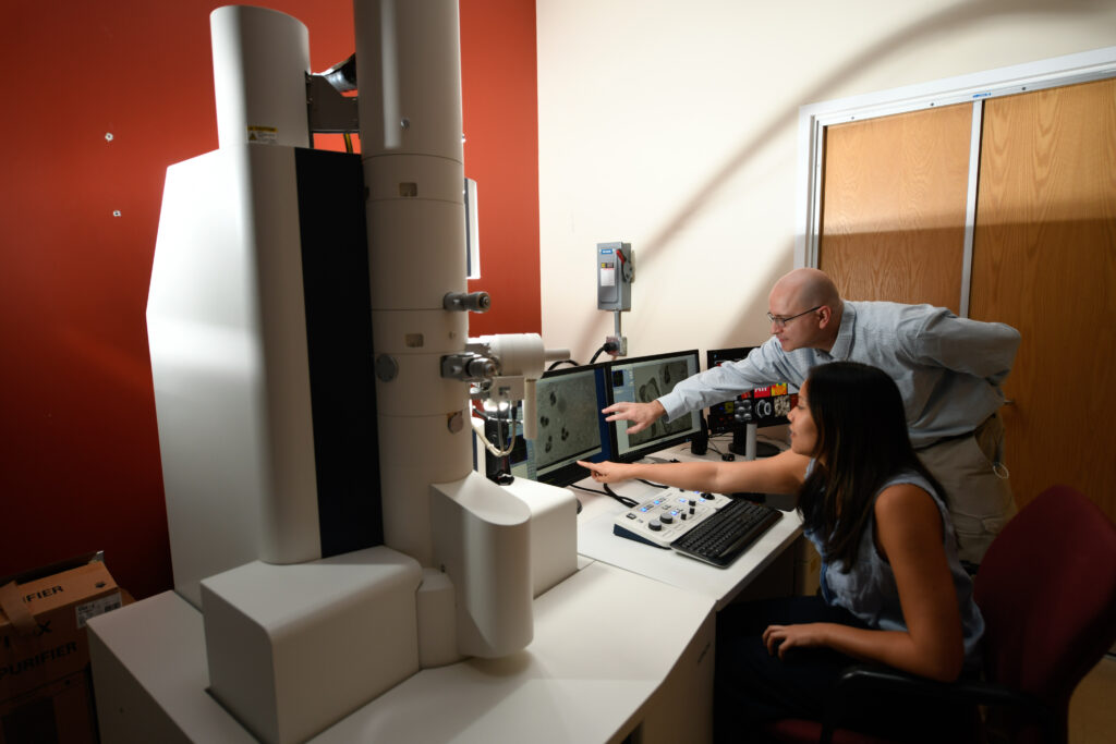
At the AIF, Dr. Bell primarily works with transmission electron microscopy (TEM), but he also does some scanning electron microscopy (SEM). The AIF primarily works with labs from the university, but they also assist external clients who want to use some of their instrumentation. Most of the samples he works with are biological, so he’s usually working in the magnification range of 50,000 to 80,000x. Through his work at the AIF, he’s also gotten to experience “many more techniques than [he] ever thought he’d work with,” such as cryo-EM. Most of the AIF’s clients bring their samples to be completely processed and imaged by the facility, but some of them will process their own samples to cut down on costs. And a few labs have even had some of their members trained on how to use the instrumentation so that they can operate it themselves. The graduate students who take this course may even be able to do their own work in the AIF after completing the course, though it can only begin to scratch the surface of what EM can really do.

“The course is really an introduction to biological imaging and electron microscopy,” Dr. Bell said. It’s mostly focused on giving students an appreciation for what EM can do, rather than providing in-depth instructions for any one aspect. The seven-week course will be split evenly between SEM, TEM, and cryo-EM. “It’ll give students a taste of what these techniques are and how they can be used for their own research interests,” he said. Students will be learning about the techniques and their association instrumentation, but they will also learn about how to prepare samples for imaging. For example, they’ll learn about how to perform critical point drying for samples they’ll be using for SEM: “It’s a much better method for drying a specimen, and it reduces cracking in the specimen [compared to air drying].”
The labs will mostly jump around between different methods and techniques, in order to ensure students are exposed to as wide a variety as possible. “There are some protocols that are multi-day processes, so we won’t be doing those,” he said, “so we’ll be focusing more on embedding and making sure students understand the theory behind EM.” Even though students won’t be able to do every single technique, they will still at least get to observe several different ones, such as ultramicrotomy, which is a way of slicing tissues into extremely thin strips. The course is open to both undergraduate and graduate students, and the graduate students are required to have an SEM project that they will work on throughout the semester. “I really like doing SEM,” Dr. Bell said, “because the sample prep is much easier and you can have an image just a few hours after you start.” The graduate students will be going to Lake Raleigh, collecting bugs, and then processing them so that they can look at them using SEM.
To see what the insides of the cells look like, and observe their organelles, you really need to use EM
Dr. Bell
The course is designed to give students a better understanding of what microscopy techniques are available to them, which ones are better for certain situations, and how they can use them for their own research. “To see what the insides of the cells look like, and observe their organelles, you really need to use EM,” Dr. Bell said. Though it’s not the focus of the course, students will also gain a better appreciation of cell biology and the details present in the world around us: “the cortex of the spider mite has these really cool ridges on it that you would never be able to see with the naked eye or with a light microscope.” The course will also be useful for students who may not have an immediate need to do EM, as these techniques are also common in medical settings, and can help students really understand the diagnostic power of EM.
Electron microscopy tends to be intimidating for students who have only heard about it through their 10th grade textbook; while it is challenging, it is also a rewarding way of experiencing the world. Students who are interested in learning more about the many types and their applications can take advantage of the BIT Program’s new course, BIT 495/595 Introduction to Biological Electron Microscopy Techniques, which will be offered in Session II of the Spring 2022 semester.
- Categories: